! STOP BENDING NOW !

Thursday 23 August 2018

Osteoporosis - Understanding some essentials

Bone health is something ignored universally but is vital in the overall health of any person.


What is Osteoporosis ?

Osteoporosis is the archetype of problems associated with old age and also the best example of a ‘silent disease’. It progresses slowly and will not be recognised till a fracture (called fragility fracture ) or disability like kyphosis or pain etc sets in.
Bone health is something ignored universally but is vital in the overall health of any person. Osteoporosis is the most common of all metabolic bone disorders. Osteoporosis literally means porous bone, is a disease in which the density and quality of bone are reduced.
It is characterized by low bone mass and microarchitectural deterioration of bone tissue, with a consequent increase in bone fragility and susceptibility to fractures .When bone loss is limited it is called osteopenia.Osteoporosis can be classified into two basic forms: primary and secondary.Primary osteoporosis (also known as involutional osteoporosis) results from cumulative bone loss as people age and undergo sex hormone changes.
Secondary osteoporosis can result from various medical conditions or diseases, or from the use of certain medications that adversely affect skeletal health. Children with specific diseases and malnutrition also can have low bone mineral density and osteoporosis.
What is the incidence of osteoporosis?
Worldwide 1 in 3 women and 1 in 5 men over 50 will experience osteoporotic fracture. In India,  estimates suggest that of the 23 crore people will be over the age of 50 years by 2015, and of that 20% are osteoporotic women.
Up to 20% of patients die in the first year following hip fractures, mostly due to pre-existing medical conditions. Less than half those who survive the hip fracture regain their previous level of function.
How is osteoporosis diagnosed?
Osteoporosis is still most commonly diagnosed at conventional radiograph. The main radiographic features of generalised osteoporosis are increased radiolucency and cortical thinning . Wedge fractures or crush fractures are seen in spine x-rays. Fragility fractures and insufficiency fractures also point to osteoporosis.
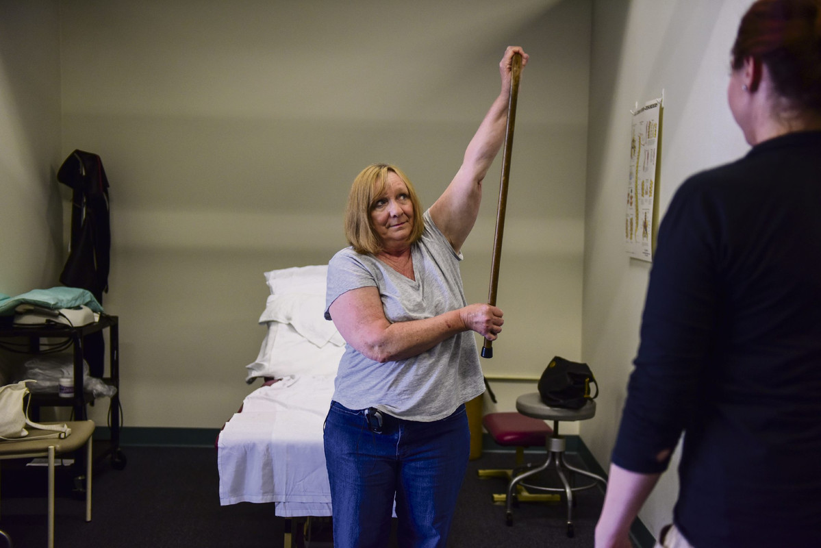
A Fragility Fracture is one resulting from any fall from a standing height or less, that results in a fracture. A bone density test is the only test that can diagnose osteoporosis before a broken bone occurs. This test helps to estimate the density of your bones and your chance of breaking a bone. A bone density test of the hip and spine by a central DXA machine is used to diagnose osteoporosis.
DXA stands for dual energy x-ray absorptiometry. Today, BMD measurements have an important role in the evaluation of patients at risk of osteoporosis and in the appropriate use of antifracture treatment.
DXA examinations have three major roles, namely the diagnosis of osteoporosis, the assessment of patients and the risk of fracture, and monitoring response to treatment. BMD measurements gives an accurate value for assessing osteoporosis and follow up.
Is osteoporosis preventable?
Osteoporosis is one condition that can be effectively prevented for vast majority if essential steps are taken. Plenty of calcium , adequate physical activity and taking in Vitamin D as appropriate are the main recommendations.
For adults ages 19 to 50 and men ages 51 to 70, the recommended dietary allowance (RDA) is 1,000 milligrams (mg) of calcium a day. The recommendation increases to 1,200 mg a day for women after age 50 and for men after age 70.Good sources of vitamin D include oily fish, such as tuna and sardines, egg yolks, and fortified milk and of course getting sunlight.Smoking, alcohol, other drug or substance abuse contribute to osteoporosis.
Treatment options
Pharmacologic treatments for osteoporosis include bisphosphonates (alendronate,  risedronate, ibandronate, zoledronic acid), peptide hormones (teriparatide which is a amino acid fragment of parathyroid hormone] and calcitonin), estrogen (in the form of menopausal hormone therapy) for postmenopausal women, and selective estrogen receptor modulators (SERMs) (raloxifene for postmenopausal women).
Who should get osteoporosis tested
For post-menopausal women below the age of 65 a bone density test is indicated if they have a risk factor for low bone mass such as low body weight, prior fracture, high risk medication use. Disease or condition associated with bone loss.
Women during the menopausal transition with clinical risk factors for fracture, such as low body weight, prior fracture, or high-risk medication use.
Men aged 70 and older. For men, less than 70 years of age a bone density test is indicated if they have a risk factor for low bone mass such as for women listed above Women aged 65 and older.


Source : Indian Express , 15th August 2018 

5 Exercises To Correct Kyphosis/Hunchback, Very Easy


Kyphosis is a condition in which the spine curved forward exceeds the normal range so that someone who experienced it will be crippled. 


About 20 to 40 percent of elderly people suffering from Kyphosis. However the current undeniable the youth had experienced it. Kyphosis generally occurs due to poor posture habits or congenital disorder since birth. 

A study published in 2009 in the International Osteoporosis find if spinal connecting exercise is done three times a week for one year are routinely able to improve posture. Here are a few sports that can be done to correct a hunchback posture. 


1. WALL SLIDES 


The first way that you can use to improve posture that humpback is to stretch the chest.

Follow these steps: 

Stand with your back upright attaches on the parallel wall. 

Give Your feet a little distance from the wall Stretch sternum by pressing your back against the wall are flat while lunges Put both palms on the wall make sure the hands outside the State Slide it slowly 

Your body position to the left of the When you shift the position of the body to the left, 

You should be able to feel the stretching that occurs in the chest and also at the shoulder 

Hold to stay in the position for a few seconds 

After shift your body to the left, then replace in the position of right Do it alternately 


2. DOORWAY STRETCH 


This movement uses the media door used as a fulcrum in a holding load of the body. 

Stand in the middle of the open door Raise Your both arms on the right and left of the door frame while you bend your elbow 90 degrees until Make this one foot forward Hold tight the door frame while pushing your body forward until you feel a stretch in your chest Hold down for 10 seconds Repeat as many as 3 sets 


3. Resistance band row 


When you to the gym, you can do this exercise using a cable machine tool help elastic. You can also use media ropes made like the workings of the machine. 


Start standing with both hands holding resistance bands Then pull the band Interesting moments of the cable machine try to position the head, shoulders, backs straight Do the way as much as 3 sets 


4. Prone Y 


The pose is similar to Cobra pose, it's just that you lift your hands to the top of the form the letter Y. Prone body on the floor or it could be use as mattress pads 

Align position of foot with the position of the shoulder After that, lift the arm until the second part of the body forming the letter Y Hold the position of the body like that for 5 to 10 seconds Do the way as much as 3 sets 


5. Upper back


One of the exercises that can be used to improve your posture is to train the upper back or the back of the top. This exercise requires the help of foam rolling so that movement could be done perfectly. 


Put the foam rolling in the spine, under the scapula Bend the knee part then let the feet touch the floor And put both hands on the back of the head in the waist Lifting slowly then begin raising her hips and lowered her hips little by little over the last 10 to 15 minutes Do the way as much as 3 sets. 


Source : Sutrisme , 15th August 2018 


Qigong Therapy for kid’s scoliosis and kyphosis


Description

I am an excellent Qigong healer. I have an amazing unique telent---figure out the root of the health problems. I have successful experiences for kid's scoliosis and kid's kyphosis. No brace, No surgery. Long term benefit. This is a very natural and powerful Chinese therapy. It's a more holistic approach, focusing on the body as a whole. I will create an unique plan that only works for your kid. After each session, I will give some different exercise that create for your kid. Your kid will have a remarkable improvement after every session. The result is better than any other traditional therapy. For more info please visit www.qigongheal.com. Thanks
Mrs. Jessica Chen Receipts for insurance SOURCE : KIJIJI   

3D prints improving accuracy of spinal surgery

New technology at one Michigan hospital allows doctors to actually see the spine before cutting the patient open.
In turn, surgeons are increasing their accuracy rate to 99% versus 95 or 96.
That improved accuracy is crucial when placing screws in the spines of people with scoliosis.
Dr. Joseph Dewitt started using this 3D technology, Firefly, a year ago.
"So having this guide, I no longer have to rely simply on visual landmarks and feel," Dewitt said. "I actually have a mechanical tool to keep everything in line"
Patients go through a pre-op scan which makes a 3D replica in the computer. 
That model is then 3D printed into the actual size to help doctors better prepare when it comes to placement and trajectory.
Even with the 3D model, the surgery can take up to 8 hours.
Recovery time depends on the case and can take anywhere from weeks to months.

More News from this link : http://www.wilx.com/content/news/New-technology-improves-safety-and-accuracy-of-spinal-procdures-490759291.html


Source : KAIT8,16th August 2018 

Irish mother forced to beg for son's life as he waits overs 14 months for spinal fusion surgery

This is simply unacceptable.

Irish mother forced to beg for son's life as he waits overs 14 months for spinal fusion surgery

In Ireland, one of the most prosperous countries in the world, a mother has been forced to beg for her child's life.
Brendan, a 13-year-old boy with scoliosis, as well as cerebral palsy, has been on the "urgent" waiting list for vital spinal fusion surgery for over 14 months.
On Thursday night, Brendan's mother Tracy took to Twitter to share photos of the increasing damage Brendan is enduring as he waits endlessly for the surgery at the Children's Hospital in Crumlin.
Tracy spoke to JOE to describe just how agonising the last 14 months have been.
"If you or I had scoliosis, that would be hard enough," Tracy said.
"But if you had no muscles, in your abdomen, your back, your sides to fight against the curve — that's Brendan. He's literally being crushed to death.
"His internal organs, his lungs, his bottom ribs are hitting the top of his pelvic bone."


"He was first placed on the urgent, I laugh, "urgent," waiting list on 1 June, 2017." Over 14 months ago.
Asked how the healthcare system justifies this waiting times, Tracy sighs hard.
"At an appointment in May, it was said that he would have his surgery in most likely July, and if everything went well, he'd be ready for the start of school this autumn" Tracy said.
"July didn't happen. I was told, it being summer, there were a lot of sports injuries, emergencies, that kind of thing, that bumped scheduled surgeries. But Brendan was never even scheduled in the first place. They just haven't put him on the schedule.
"We need to ask why and demand different. Something's gone wrong, obviously. It's massively dysfunctional, massively so. It's inhumane. This is neglect.
"He's a severely disabled child, you can't get more vulnerable in society, than a severely disabled, non-verbal child who is 100% dependent on every single care in his life, 24/7. And this is how he's been treated. And that is an indictment on those that have the power to make things different."


On Minister for Health Simon Harris, Tracy says he won't reply to her emails directly. On Minister for Disability Issues, Finian McGrath, Tracy says he has not replied once to her, even though he has met Brendan before.
Tracy is now waiting on the outcome of a special meeting that has been called at the hospital to discuss Brendan's case, after 14 months of waiting.
"It wouldn't have happened without the pressure. I would still be waiting. If I hadn't put the pressure on directly, which I hate doing, nobody should have to do it. Posting pictures of their child for the public to see, especially in a vulnerable and painful situation. God. No parent wants to do that. But it seems to be the only way to get them to do something that they should be doing in the first place."
Tracy notes that the compression of Brendan's body has begun to force wounds to open — which would disqualify him from surgery. This means that by waiting so long to act, the healthcare system may have made it even harder to operate on Brendan.
Drastic waiting lists have become par for the course in the Irish public healthcare system. "Over 14 months on an urgent waiting list," Tracy says. "That doesn't make sense."
"That's part of the problem. We need to not be used to it. We can't wait until it affects someone we love. We all have to be outraged by it for it to change."
There is currently a Go Fund Me page to help Tracy pay for suitable accommodation for Brendan, which is almost halfway towards its goal.
"Brendan requires a properly equipped, suitably modified bungalow for his lone-parent mother to safely continue her full time 24/7 care of him, especially when that operation is completed," the Go Fund Me page says.
"Brendan, his younger brother Declan, and their mum Tracy currently live in a rental home that doesn’t meet those requirements. There is no suitable social housing available and the wait list is years long."
UPDATE: Tracy has confirmed that Brendan has been given a tentative date of October 8 for the spinal fusion survey, as a direct result of the social media pressure applied over the last 24 hours.
This will partially depend on an anaesthesia review which will take place on Monday.


Source : JOE , 17 August 2018 

A novel approach to complex spinal surgeries: The successful use of 3D-virtual and 3D-printed models

3D
A 3D-printed spine model with 3D-printed osteotomy templates (red). 
A novel approach reported by Peter Pijpker and colleagues from the University Medical Center Groningen, Groningen, The Netherlands, outlines the use of 3D virtual planning and 3D-printed models that have the ability to change how complex spinal surgeries are managed. Using this technique, surgeons were able to successfully reduce severe kyphosis in a young girl who suffers from skeletal dysplasia.
Pijpker et al emphasise the need for more precise methods of surgical planning. They state that a major advantage regarding the use of 3D virtual planning is the ability for the preoperative plan to correlate precisely with the actual operation: “This technology can give rise to new possibilities for the surgical planning of spinal deformities,” they wrote.
For patients with thoracic kyphosis, vertebral column resections and pedicle subtraction osteotomies (PSO) are widely indicated. To reduce the risk of injuries occurring during the osteotomy and pedicle screw insertion, computer assisted surgery (CAS) systems are commonly used. However, the procedure remains technically demanding with a risk of major complications.
Furthermore, the authors note that recent research in relation to individualised templating for spine surgery is currently limited to drill guides for accurate pedicle screw placement. Although patient-specific osteotomy templates have been described for knee arthroplasty, the authors report that this is the first paper describing this technique for complex spinal osteotomies.
The report describes the case of a young girl with severe angular thoracolumbar kyphoscoliosis.  A closing wedge extended pedicle subtraction osteotomy was first virtually planned using medical computer software. Individualised osteotomy guided templates were then designed for the translation of the planned 3D-wedge towards the surgical procedure.
The authors note that the 3D planning and templates facilitated surgery in various ways. Not only did it allow for a 3D insight into case-specific anatomy and identification of vertebrae levels (as well as malformed vertebrae visualisation) during surgery, but it enabled the direct translation of the planned pedicle subtraction osteotomy into the surgical site using 3D printed individualised osteotomy guided templates. Additionally, the surgeons reported that studying the 3D anatomy in a multidisciplinary team facilitated the surgical procedure due to enhanced spatial orientation.
Speaking to Spinal News International, Pijpker comments: “In these severe kyphoscoliosis cases and other high risk or highly complex spinal procedures, these 3D models help our teams to train and prepare. The templates may, together with patient specific drill guides (another topic we are currently working on), contribute to safer spinal procedures. We believe that the 3D planning and printing technology is going to transform the way complex surgery is managed, whether through onscreen 3D visualisations, patient-specific bone models, individualised guiding templates or custom-made prosthetics.”
In relation to the case, the post-operative period was uneventful and the patient was discharged without any neurological deficit after eight days. The early postoperative X-rays showed a satisfactory correction of the kyphoscoliosis, as the kyphosis angle was reduced from 74° to 22° and her coronal plane was normalised.
Through the successful correction of the kyphoscoliosis in this case, it is clear that the novel use of 3D-virtual planning, 3D-printed spine models and osteotomy guiding templates have facilitated the performance of the osteotomy. In the report, the authors also note that this technique is feasible for the surgery of complex spinal deformities. They write that, from the surgeon’s perspective: “The templates and bone models provide valuable guidance during the osteotomy in the severely deformed anatomy.”
The authors reiterate that this method of spinal correction can be cost-effective, while it may help to reduce surgery time. Furthermore, it may preclude the need for intraoperative radiography—especially when combined with patient drill guides.
However, while the templates provided great directional support for the surgeon, the authors note that the use was nevertheless limited to the first stages of pedicle subtraction osteotomy. For example, when approaching the apex of the wedge, temporary rods had to be placed for stabilisation and safety.
As this is a novel technique, future designs of templates can be optimised by incorporating inlets for rod-positioning. Regarding the future of this method in terms of feasibility, the authors said that current feasibility was limited to describing the technical aspects, as well as the use of this new method in a qualitative manner. Therefore, to move forward with this technique, a comprehensive accuracy study is needed in relation to the clinical outcome; assessing its efficacy in a quantitative manner.
Nonetheless, the authors conclude by saying that, in addition to contributing to safer spinal osteotomy procedures, the novel use of this workflow involving 3D-virtual and 3D-printed models may change how complex surgeries are managed.

Source : Spinal News International , 17th August 2018 


Sparrow Hospital improves scoliosis surgery through 3D modeling

Lansing, Mich.-based Sparrow Hospital implemented a 3D printer to improve outcomes of scoliosis surgery patients, according to WKAR.
Before a patient's scoliosis surgery, physicians use the 3D printer to make a model of the patient's spine to practice the procedure and determine the proper entry point for the hardware to properly address spinal curvature.
 "We take a CT scan, we're able to load that CT scan into the computer, 3D model it, and then at every level that we determine that we need to fuse, we're able to create a patient-specific guide for that level, " Joseph DeWitt, MD, a pediatric orthopedic surgeon at Sparrow Hospital and McLaren Greater Lansing Hospital, told WKAR. "This allows us to very, very accurately and safely place our hardware, which consists primarily of screws, a couple of hooks, and then rods. "
Dr. DeWitt has completed 20 surgeries using the 3D modeling technology.


Source :Becker Hospital Review , 21 August 2018 

Saturday 11 August 2018

Nashville boy battling very rare condition gets Titan support


Kayden Ivey (Photo/Brittney Ivey)
The Tennessee Titans helped shine a light on a rare disease a Nashville boy is tackling daily.
Kayden Ivey has CLOVES syndrome. CLOVES Syndrome stands for Congenital, Lipomatous, Overgrowth, Vascular Malformations, Epidermal Nevi and Spinal/Skeletal Anomalies and/or Scoliosis. According to the CLOVES Syndrome Community, it has been diagnosed in about two hundred people so far.
The Titans know the value of teamwork and the importance of National CLOVES Syndrome Awareness Day. On Friday, the NFL team invited to their practice; the only kid in Nashville and the state with the rare condition.
Kayden Ivey says, "It's really fun to have CLOVES day because it tells you: you're not the only one, you're not alone. Some people don't have what I have, so when I tell people they don't know what it is."
Given the recent discovery of this syndrome in 2009, there are likely many more people who remain undiagnosed. People living with cloves often experience neurological complications, pain, and significant and aggressive overgrowth of affected parts of the body.
"My right leg is longer than my left leg. And my hands are bigger than what you expect to be on an 11-year-old," Kayden explains.
The 11-year-old was diagnosed in Cincinnati at age: 6, according to his mother, Brittany Ivey.
"I went everywhere because no one knew what was wrong with my baby. He used to get these big tumors on his back and he's been diagnosed with cancer so many times," Kayden's mom explains,
"I even had a doctor one time tell me: 'enjoy Kayden because honestly if we ever get the money or funding to produce or research, or get a cure for Kayden...Kayden won't get it. It will go to cancer. He's one, it's a million other people.' That's why CLOVES Day is so important to me because it's just him, but don't forget about it....don't forget about my baby."


Source : Nashvile , 5th August 2018 

Spinal fusion surgery changed this Greensburg woman's life

Charlene Rose wanted her mobility back.
For two years, the 65-year-old Greensburg native grappled with debilitating neck and head pain that seemed to be getting worse.
Every time she turned her head even slightly to the left, a sudden jolt that felt like an electric shock reverberated from her upper spine to the top of her head. An earache throbbed day and night.
Known for her impeccable homemaking skills — and formerly running a medical facility cleaning company — Rose hated that she couldn't carry out daily chores. She struggled to clean and cook. She could no longer safely drive. Rose only avoided the painful shocks by tilting her head awkwardly to the right and holding it there.
"I had to walk around with my head tilted. I could never straighten my head. I couldn't work like that," recalled Rose, who reluctantly quit her job at Walmart this past spring.
Physical therapy alone wasn't working, and Rose was getting desperate. She finally found a doctor who said he could help — Dr. Kevin Walsh, a neurosurgeon who runs the Adult Spinal Deformity Center at Forbes Hospital in Monroeville.
Walsh proposed a pair of surgeries, to be performed back to back, to correct cervical spine deformities and replace Rose's degenerated vertebrae with strong cadaver bones via a spinal fusion. If his plan worked, Rose likely still would live with some level of chronic discomfort, but she would be freed from the sudden shocks and never-ending headaches and earaches.
The catch: recovery would be long and agonizing.
"I'm going to have to cause you pain to heal your pain," Rose recalled Walsh telling her.
She decided to go for it.
What she didn't anticipate was just how excruciating the recovery would be and how reliant she'd become on her 79-year-old husband, Joe. And she definitely didn't foresee how frightening it would feel to confront a series of unexpected complications.
Charlene Rose, 65, of Salem works with physical therapist Molly Bruggeman at Excela Health Outpatient Rehabilitation, in Delmont on Monday, July 23, 2018.

THE SURGERY
More than 200,000 patients undergo cervical spine surgeries annually, the American Association of Neurological Surgeons reports. Typically, the procedure is reserved for intolerable pain that can't be cured otherwise. Rose presented a challenging case because she had compounding issues — a degenerative disc disease as well as a spinal deformity caused by kyphosis, an inward curvature of the spine that forces the patient's head forward.
Treating her would require two surgeries in one day, so Walsh could cut through both the front and back of her neck.
On the morning of May 21, Rose lay on her back under anesthesia inside an operating room at Forbes Hospital. Walsh and his team surrounded her.
Surgical tools are laid out inside an operating room at Forbes Hospital in Monroeville on May 21, 2018.

An X-ray of Rose's spine mounted along a wall made visibly clear what was wrong — the insides of three vertebrae looked deteriorated, appearing gray and black and crumbling in comparison to the rest of the solid white, healthy bones lining her spinal cord.
Using a surgical robot and $500,000 microscope, Walsh patiently scooped out the degenerated vertebrae pieces before screwing in healthy replacement bones from cadavers.
About five hours in, Walsh closed the first incision. A few more medical workers hustled into the OR to prep the for "the flip" — a move that saves time and improves safety by using the same specially equipped, $120,000 operating table without having to move the patient.
The double-surgery was expected to take 10 to 12 hours. But 14 hours after the surgery started, Walsh told Rose's husband they needed more time.
Surgeons work inside an operating room at Forbes Hospital in Monroeville on May 21, 2018.

There had been a "mishap" with a piece of equipment during the surgery, Walsh said. When Rose woke the next morning, she wondered what had happened to her very sore and stitched-up head.
"You were operating on my neck," she recalled telling Walsh. "How could there be a hole in my head?" A nurse explained a clamp that holds the head during surgery slipped, causing a small laceration on the back of her head.
"It is a rare occurrence, but it does happen, especially with the three-point head holder that we have to use for this type of positioning," Walsh later told the Tribune-Review.
The U.S. Food and Drug Administration has warned about the risks of using skull clamp systems, citing more than 1,000 medical device reports resulting in more than 700 injuries — skull fractures, deep cuts, bruises and blood clots — associated with slippage or movement of the clamp before or during surgeries between January 2009 to 2016.
Still, the FDA concludes that the benefits of a skull clamp outweigh its risks — surgeries like Rose's likely couldn't take place without one. Allegheny Health removed the clamp involved and is investigating the incident.
Surgeons work inside an operating room at Forbes Hospital in Monroeville on May 21, 2018.


The unexpected head wound aside, what Rose thought would be a two- to three-day hospital stay turned into a few days in the hospital followed by several more in a nursing home. Then, less than three weeks after the surgery, Rose woke alarmed to find her head and pillowcase soaked in blood. Her wound oozed with crusty, yellowish fluids. A blood culture showed she had a bacterial infection. She'd have to go under the knife again. Complications tend to happen in 20 to 45 percent of similar types of cervical spine operations, and roughly half the time the problem is a wound issue like Rose had in which the incision hadn't closed properly, according to Walsh.
"It's very scary," said Rose. "You don't know if they're going to be able to get it (the infection) all out."
The follow-up surgery in mid-June went smoothly — but the intensity of the pain that followed began to make Rose question the whole thing.
Charlene Rose (left), 65, and her husband, Joe, of Salem are seen reflected in a mirror at their home while talking to reporters about the second surgery she had to go through to clean out an infection on Tuesday, July 3, 2018.

THE RECOVERY
During the first few weeks post-surgery, Rose had trouble sleeping more than a couple hours at a time and often woke to violent stabs of pain.
"When I first came home, I couldn't stand it," Rose said. "Every single muscle and bone in your neck, it was like somebody was stabbing it with a knife.
"I couldn't do anything by myself."
Each morning, her husband carefully helped her out of bed and fixed her breakfast, usually scrambled eggs and toast or toast and coffee, before she took her medications.
He washed her hair and laid out three outfit options before helping her into the one she liked best.
Her oldest daughter came by to clean, and neighbors sent over homemade chicken and vegetable beef soups.
Rose described the pain as "10 times worse" than the electric shocks, with every minor move she made tugging at the long bands of sutures connecting layers of muscles, nerves and skin across her back and down her spine. She said it even felt worse than the time in her late 20s when she and her husband hit a log while riding a snowmobile, throwing Rose into the air and crushing and compressing several vertebrae.
Charlene Rose, 65, of Salem works with physical therapist Molly Bruggeman at Excela Health Outpatient Rehabilitation in Delmont on Monday, July 23, 2018.


She spent seven months with a board strapped to her back, during which she learned she was pregnant. After relearning to stand, she had to walk at least a mile a day to gain strength in the weeks leading up to labor — and she ended up giving birth to her daughter at home before medics arrived.
None of that compared to her post-cervical spine fusion surgery, she said. "I breezed through childbirth," Rose said with a chuckle. "Even when I broke my back, it was nothing like this."
As recently as two weeks ago, Rose thought she might regret the surgery altogether. But, gradually, the pain began to subside.
She weaned off opioid painkillers and got down to taking only Tylenol and her antibiotics.
She started driving again, making short trips to the supermarket so she could whip up homemade treats like pierogis and pineapple upside down cake.
Last week, during her fifth physical therapy session, Rose boasted, "I feel wonderful."
"I'm mobile now," Rose said cheerfully as she performed arm stretches using bungee cords at Excela's physical therapy office in Delmont.
"I do my cooking, and I can clean up the house and make the beds and wash the clothes," she said. "My scars are healing nicely, and it gets a little less painful each day."
Walsh said that Rose "will most likely have some neck pain for the remainder of her life, but if she's already 50 percent better than she was before the surgery, that's a good indicator that she's going to keep improving over time." He anticipated that Rose's full recovery could take three months to a year.
"I told 'em that whenever I can curl my hair," Rose said as she grazed the ends of her chin-length blonde locks with her fingertips, "then I'll know I'm fully healed."


Source : Trib Live , 5th August 2018